



From ancient Egyptian medical records to mammography and emerging breath-analysis research in Lebanon, the history of breast cancer detection reflects a continuing pursuit of earlier, less invasive and more accessible diagnosis.
Breast cancer detection enters a new era

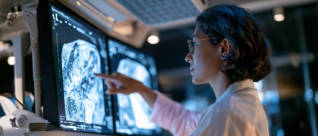
The earliest record of breast cancer is among medicine’s oldest documents. Evidence of the disease appears in ancient Egypt, around 3000 to 2500 B.C.E. For millennia afterward, detection meant a hand finding a lump, usually late and often too late.
The surgical era sharpened the stakes without improving the odds. William Halsted performed the first successful radical mastectomy in 1882 at Johns Hopkins Hospital, and his theory of stepwise spread from breast to nodes to the body at large gave early detection its rationale, though doubts about the approach lingered for generations. If a tumor could be caught before it travelled, the logic ran, it could be cured.
The tool for catching it arrived from physics. In 1913, the German surgeon Albert Salomon X-rayed some 3,000 mastectomy specimens, found visible differences between cancerous and normal tissue, and noted the significance of microcalcifications. Yet his images showed removed tissue, not living patients, so clinical use did not follow. Not until 1927 did Otto Kleinschmidt publish what is regarded as the first authentic mammography, crediting his mentor Erwin Payr.
Acceptance came slowly. Widespread screening use followed a 1966 study led by the American radiologist Philip Strax, and randomized trials, beginning with the Health Insurance Plan study, supplied evidence of reduced mortality. Women who attend mammography screening show roughly a 30 percent reduction in breast cancer mortality compared with those who do not. Digital imaging, tomosynthesis and contrast-enhanced techniques have since refined the picture, while researchers pursue blood-based approaches such as liquid biopsy and circulating tumor cells.
Yet the method retains familiar burdens, repeated radiation, false alarms, biopsies, and the long wait for a verdict.
The Beirut laboratory with a breakthrough
Dr. Amer Sakr, Vice President for Academic Research at the American University of Science and Technology (AUST) and Director of its Faculty of Health Sciences, is leading a research program into breath analysis. His starting point is the biology of the tumor cell itself.
“Cancer cells have recently been described as cells that use fermentation,” he told The Beiruter, likening their handling of sugar to winemaking, where microorganisms convert sugar to alcohol without oxygen. Healthy cells, by contrast, burn nutrients with oxygen. The distinction matters because the by-products differ. “As a result, they release different types of volatile organic compounds, or VOCs,” Dr. Sakr noted. Those compounds enter the bloodstream and, in the lungs, “can pass into the air and be released through breathing.”
The ambition follows directly. “The idea is to monitor these VOCs and determine, by analyzing a person’s breath, whether cancer may be present.” Some compounds, he observed, “may be present at higher levels when cancer is present and at lower levels when it is absent, while others may show the opposite pattern.”
The project is a collaboration with the American University of Beirut Medical Center, and it has already widened in scope. We are currently working in collaboration with Dr. Ahmad Bulbul and Dr. Naji Saghir, who is responsible for breast cancer care and serves as the hospital's chief oncologist,” Dr Sakr explained. An infectious disease specialist is working with the team to see whether lung infections could be detected the same way, potentially bypassing laboratory tests that are costly and slow.
For breast care, Dr. Sakr sees a gentler pathway. “When we can detect cancer through breath analysis, women do not need to undergo mammography as frequently as they do now,” he offered, pointing to cumulative radiation exposure and to the anxiety and expense of biopsies that sometimes prove unnecessary. A breath test could be repeated according to individual risk: “For someone with a family history of breast cancer, testing might be considered every six months, for example.” He was careful to add that these are “examples of how the technology could be used, rather than established screening recommendations.” He believes the same principle could extend to prostate cancer, where PSA results can mislead in both directions.
He is keen to place the effort within a wider scientific movement rather than present it as an isolated venture. “This type of work is progressing in countries such as China, India and Russia, as well as in Europe, where interest is growing,” he stressed. The implication is that breath-based diagnostics are no longer a fringe curiosity, and that Lebanon has a chance to take part rather than watch from the sidelines.
He framed the work as proof that Lebanon need not wait on others. Dr Sakr, Lebanese-American scientist who spent 15 years in the United States after moving at 16, and who holds a U.S. patent, he recalled asking himself: “If this technology can be developed abroad, why should we not develop it here?” Early results, he reported, have been “very promising”, clinicians send samples, and the analysis takes “approximately one hour.”
Whom would it serve first? “Anyone with a family history of breast cancer should consider being tested, regardless of age,” he stressed, describing a baseline measurement followed by monitoring for rising markers. The same approach, he added, could track chemotherapy response and watch for recurrence.
Each leap in detection began as an idea in a laboratory awaiting proof, all with one dream in common, to make detection earlier, less of a burden and more accessible, giving patients a better chance of confronting breast cancer before it advances.
-
-

-
Jenna GeageaReporter/Writer
-
